Isolates


General information
Name: Rhizophagus intraradices
Mycorriza Bank #: Location Sate Country Latitude / Longitude
CMCC/AM-1106 Mukteswar Uttarakhand India 29.4604° N, 79.6558° E
Classification and associated taxa
Kingdom: Fungi
Division: Glomeromycota
Class: Glomeromycetes
Order: Glomerales
Family: Glomeraceae
Genus: Rhizophagus
Biochemical information
Fatty acid methyl esters (FAME) C-12,C-14,C-16,C-16:1,C-18,C-18:1
Functional information
Percentage enhancement of morphological parameters as compared with un-inoculated control on Lycopersicon esculentum (tomato)
Shoot Length Number of branches Shoot fresh weight Shoot dry weight
CMCC/AM-1106 2 25.3 4.5 5
Percentage enhancement of macro nutrient parameters as compared with un-inoculated control on Lycopersicon esculentum (tomato)
Nitrogen Phosphorus Potassium
CMCC/AM-1106 0.62 170.27 116.50
Percentage enhancement of micro nutrient parameters with as compared with un-inoculated control on Lycopersicon esculentum (tomato)
Iron Manganese Zinc Copper
CMCC/AM-1106 425.82 516.99 NS NS
Biocontrol efficacy *
Pathogen Fusarium oxysporum Rhizoctonia solani
CMCC/AM-1106 22.50 18.33
NS= Not significant
Spores of Rhizophagus intraradices

Shape: The shape varies from globose, sub-globose, and sometimes irregular, with many spores elliptical.

Color: The color of spores ranged from white.pale cream to ellow brown and sometimes light brown.Spores shows golden yellow color in PVLG and golden yellow to golden red in Melzer's:PVLG.

Average diameter: 110-(120)-180 μm

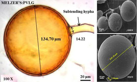
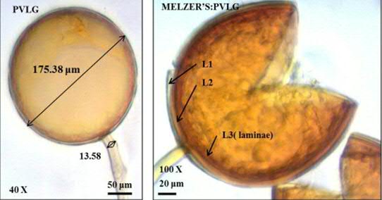
Spore after reaction with Melzer's Reagent

Spore wall layer 1: L1: Outermost layer, hyaline, mucilaginous, 0.5-3.5 μm thick, staining pinkish red to pale purple in Melzer's reagent when intact in juvenile spores. With age, this layer almost always degrades and decomposes naturally after which it appears granular and may accumulate some debris.

L2: Adherent to the mucilaginous outer layer, hyaline, 1.5-4.9 μm thick when intact in young spores. With age, this layer degrades concomitant with L1 and also acquires a granular appearance or sloughs in patches. Mature spores often lack both L1 and L2 or they are present together as rough patches.

L3: A layer in that it consists of pale yellow-brown sublayers (or laminae) that either remain adherent or separate with applied pressure. Degree of separation among sublayers varies considerably among spores and often is affected by age, amount of parasitism, or amount of applied pressure after mounting. Thickness varies from 3.2-12 μm in mature spores. This layer forms simultaneously in the wall of the subtending hypha.